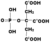
Figure 2. The structure of phosphocitric acid.
Crystal growth and its inhibition related to biological systems, (biomineralization, anticalcification therapies for pathological calcium crystal deposition diseases)
Calcium-containing crystal deposition diseases represent a group of clinically heterogenous calcific diseases that are a significant source of morbidity. Phosphorylated carboxylic acids are powerful inhibitors of biological crystallization as it relates to those diseases. Phosphocitrate (PC), a naturally occurring compound, is particularly potent. It has been demonstrated to inhibit the transformation of calcium phosphate to hydroxyapatite (Ca5(PO4)3(OH)), the deposition of calcium oxalates, the crystallization of octacalcium phosphate (Ca8(HPO4)2(PO4)4�5H2O), and calcium pyrophosphate (Ca2P2O7�2H2O), and the formation of struvite (Mg(NH4)(PO4)�6H2O) in vivo. Overall then, the compound attracts keen interest from the viewpoint of its non-toxic nature and potential to influence biomineralization in many diverse biological fields.
This project aims toward discovering more efficient calcification inhibitors based on phosphocitric acid and other related molecules. Thus far, we established that a mixed Ca/Na salt of phosphocitric acid causes an increase of it plaque growth activity from 30 % (for "plain" phosphocitrate) to 95 %. These studies were done on rats and the growth of an induced plaque (mainly hydroxyapatite) was followed.
|
Figure 2. The structure of phosphocitric acid. |
Our research program on modeling of biomineralization processes focuses on a number of topics. These include:
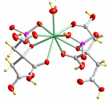 |
Figure 3. The structure of the mixed Ca/Na salt of phosphocitric acid. |
Recent experiments on combating spontaneous osteoarthritis in Guinnea Pigs showed that the complex CaNaPC showed enhanced efficiency as pathological biomineralization inhibitor compared to that of NaPC (Figure 4).
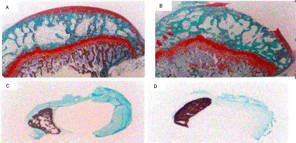 |
Figure 4. (a) Histology of 6- month old guinea pig tibia plateau after treatment with CaNaPC (40mg/wk) for 2 months. (b) Histology of untreated control 6-month old guinea pig tibial plateau. (c) Xcross-section of meniscus of 6-month old guinea treated with CaNaPC (40 mg/kg//wk) for 2 months. Note most of the calcified deposits (dark brown color) were resorbed. (d) Xcross section of meniscus of untreated control 6-month guinea pig. |